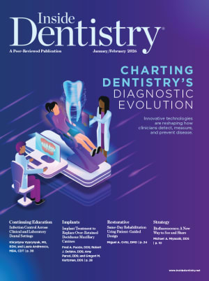
Diagnostic dentistry is undergoing a rapid transformation. Tools that once relied heavily on tactile judgment, two-dimensional imaging, and subjective interpretation are now being supplemented by artificial intelligence, multimodal imaging, salivary and genetic assays, and engineering-based structural measurements. Together, these technologies are expanding what clinicians can detect early, measure objectively, and communicate clearly, and revealing a profession moving toward personalized, preventive, data-driven care.
AI, Imaging, and the Shift Toward Early Detection
The integration of artificial intelligence (AI) into dental imaging is one of the most profound changes described by experts. AI offers exponential gains in the visibility and interpretability of radiographic information.
Lou Graham, DDS, who has spent over 40 years in clinical dentistry as well as teaching at the University of Chicago and internationally, guides practitioners toward ways to extend the life of a tooth through careful diagnostics and minimally invasive treatments. “My approach has always been very conservative-based,” he says, contrasting it with the “crown-happy” culture in dentistry. Dr. Graham notes that once enamel is removed, it’s gone for good, and teeth often enter a cascade of restorative procedures that could have been avoided with early intervention. His career now centers on clinical education and evaluating developing technologies that are reshaping oral care.
This approach also informs his overall philosophy of dentistry. When asked what he loves most about his chosen profession, Dr. Graham says, “I love the relationships that I have continued to build throughout my life. They really become extensions almost of my family.” While creating a beautiful smile or a successful implant case is gratifying, he says, it’s the long-term trust with patients that he finds most rewarding. His goal, he says, is to get his patients’ teeth to their 75th birthdays.
Improving diagnostics, Dr. Graham explains, is essential to this pursuit. He is highly optimistic about the new tools. For decades, he points out, dentists have relied on explorers and 2D x-rays, which he says lead to informed guesswork. “An explorer detecting occlusal decay is at best 50%, and a 2D x-ray evaluating interproximal decay is 60% at best,” he points out. He notes that dentists and students alike have often encountered caries that appear minor on x-ray but turn out to be much larger clinically. “Now we can minimize the guessing,” Dr. Graham says, predicting that within five years, AI will also provide treatment plans, assist with patient communication, and even manage insurance billing.
“If we really want to escalate our minimally invasive dentistry, we have to diagnose earlier and better to be more preventive,” Dr. Graham says. AI, he says, is transforming this landscape. He points out that algorithms can read 500 to 800 shades of gray on an x-ray, far beyond the 30 to 50 shades detectable by the human eye, allowing dentists to routinely identify early lesions, calculus, and incipient caries with far greater precision. This marks a revolutionary shift in how we manage non-cavitated lesions, allowing for far greater accuracy and reducing the need for drilling. This transformation is made possible by AI combined with emerging treatments like vVARDIS Curodont Repair Fluoride Plus, which reduce or eliminate the need for drilling, he says.
As Dr. Graham’s insights highlight the foundational shift AI brings to radiography, other clinicians describe how these tools influence accuracy, consistency, and team performance.
Thomas J. Giacobbi, DDS, a practicing clinician with more than 30 years of experience and the host of the Dr. Tom Show podcast, believes that emerging diagnostic technologies are reshaping dentistry in ways that directly influence clinical decision-making and team development. He notes that among the current tools driving meaningful change, AI applied to radiography stands out as one of the most impactful. Dr. Giacobbi explains that AI not only supports diagnostic consistency but also has the potential to enhance team performance by evaluating image quality over time. He says, “Imagine that this technology could provide feedback on image quality based on the team member taking the x-rays.” By identifying patterns such as recurring overlaps or missed apices, AI could highlight opportunities for targeted training and improved technique.
Dr. Giacobbi also points to salivary diagnostics as a rapidly evolving category that is expanding the scope of what clinicians can learn from noninvasive testing. While earlier methods focused on simple measures like salivary pH, today’s diagnostics are far more sophisticated and align well with patient interest in personalized care, Dr. Giacobbi says. “Salivary diagnostics are minimally invasive, affordable, and more sophisticated than ever before,” he says. For practices looking to expand their diagnostic capabilities without significantly increasing chair time, these tests represent an accessible and patient-friendly option.
When considering how these innovations may influence workflows, Dr. Giacobbi emphasizes that AI-driven radiographic analysis already benefits both clinicians and insurance processes. He notes that AI tools can provide estimates of lesion penetration in dentin or measurements from CEJ to crestal bone, which can help illustrate disease progression clearly and objectively. He also underscores the value of pairing longstanding tools such as intraoral cameras with AI-annotated radiographs to strengthen patient education and build trust in the diagnostic process. These combined visual aids, Dr. Giacobbi says, help patients better understand conditions that cannot be seen in a standard photograph.
Peter Auster, DMD, FICD, FACD, a clinician with more than 30 years of experience who teaches dentists nationally and internationally and has served as founder and past president of the Greater New York Academy of Cosmetic Dentistry, situates the current technological transition within a broader digital evolution. He views today’s rapid evolution in diagnostic technology as a pivotal moment for the profession, driven in large part by advances in AI.
Dr. Auster believes that AI presents meaningful opportunities as long as clinicians remain in control of diagnosis. He explains that dentistry is evolving faster than ever before and that AI, when used responsibly, represents a huge advancement. He points to its growing role in radiography and CT scan interpretation as well as its expanding influence in practice management. He also notes that digital tools such as scanning systems have relied on AI for years for functions like stitching, and that “the latest generation of scanners and 3D printers takes our dentistry to the next level with no sign of letting up.”
Dr. Auster is a strong proponent of AI radiography and has firsthand experience in integrating these tools into clinical workflows. He uses several programs, including Pearl, and says that AI “totally changes an office, from diagnosis to treatment planning to far more useful morning huddles.” He emphasizes that AI’s value becomes clear when clinicians view it as a supportive aid rather than a replacement for clinical judgment. To him, the key is recognizing that AI can produce false positives, false negatives, and limitations tied to poor image quality, making the dentist’s oversight essential.
Dr. Auster stresses that high-quality radiographs and strong foundational training remain central to effective diagnosis, especially for newer clinicians. “Young dentists still need to learn how to diagnose,” he notes, adding that AI can assist that process when paired with proper instruction. For Dr. Auster, the promise of AI and digital dentistry lies in their ability to elevate clinical efficiency and accuracy without diminishing the clinician’s role—enhancing, rather than replacing, the skills that define high-quality dental care.
Microbiology, Genetics, and Noninvasive Biological Diagnostics
Beyond enhanced imaging, several experts highlight the rapid expansion of not only salivary testing but also genetic assays that identify patients’ underlying biological risk factors well before any clinical signs emerge. Dr. Graham is excited about genetic testing. AI Genetics, a company based in Canada, offers a swab that can help predict a patient’s caries and periodontal risk—low, intermediate, or high—years before disease onset. He pairs this with saliva testing to monitor bacteria, matrix metalloproteinases, and other biomarkers, which can track whether a patient is improving or requires more preventative care.
Other technologies Dr. Graham highlights include transillumination and fluorescence. He’s been using DEXIS CariVu since 2014 to see cracks, craze lines, and proximal lesions without x-rays. He notes that the KaVo DIAGNOcam Vision Full HD combines fluorescence, transillumination, and a high-definition camera to enhance early detection. CaviSense, a single-use interproximal device, evaluates demineralization through pH changes, while GC Tri Plaque ID Gel highlights plaque and cariogenic risk in three ways, helping patients visualize their oral hygiene. Even home care has advanced. Dr. Graham praises the Slate power flosser as an effective tool for patients who struggle with traditional flossing.
Dr. Graham’s teaching today reflects a broad perspective. His focus has shifted to national speaking and hands-on education delivered through Catapult Education, the company he co-founded, now featuring more than 30 speakers. Most of the companies and technologies he references in his work partner with Catapult, underscoring the organization’s role in bringing promising innovations to clinicians.
When it comes to periodontal care and oral-systemic connections, new tools are emerging as important pillars of diagnosis. Periodontist Sam Shamardi, DMD, an educator, author, and entrepreneur, recently returned to Southern California after more than 16 years practicing on the East Coast. Alongside his clinical work, he has spent years in academia, serving on the Harvard postgraduate faculty and as an adjunct assistant professor at the University of Pennsylvania. He is also focusing on an often-overlooked issue in dentistry: hearing loss among dental professionals. When it comes to diagnostics as it relates to periodontology, he says, “With perio, we truly have the ability to help the patient, not just with their oral health, but their systemic health.” He points out that periodontics sits at the intersection of dentistry and medicine, but that this connection remains underappreciated both within and outside the profession.
“So much of what we sometimes miss comes as a result of either not having all the tools at our disposal or being rushed in the chair,” Dr. Shamardi says. He highlights three technologies that are transforming periodontal diagnostics: AI, saliva testing, and ultrasound. “AI helps us identify areas of bone loss, periapical lucencies, calculus, and infection that the naked eye might miss,” he says, noting that compared to traditional radiographs, AI software can distinguish vastly more gradations of gray, pinpointing areas that may need closer inspection. “Its job is not to diagnose; its job is to simply point out areas that may need closer inspection,” he says.
Saliva testing, Dr. Shamardi says, reinforces the link between oral and systemic health. “We can now identify five specific bacteria that have a direct impact on periodontal disease and implant complications, but also on systemic conditions like Alzheimer’s, diabetes, cancer, and adverse pregnancy outcomes.” With this knowledge, clinicians can tailor treatment plans and adjust long-term maintenance strategies. He says it’s particularly valuable for implant patients because it can flag those who are at higher risk for developing peri-implantitis.
Ultrasound is another emerging tool, Dr. Shamardi says. “We can measure pockets, identify defects in bone or tissue, and even assess blood flow after procedures like gum grafts to evaluate healing and implant protection,” he says.
Shervin Molayem, DDS, a periodontist and implant surgeon practicing in Los Angeles, emphasizes the critical role of technology in making diagnostic processes in dentistry more consistent and precise. With more than 15 years in clinical practice and a track record of innovation, he has focused on integrating multiple diagnostic tools to create objective, reliable measures for patient care.
Dr. Molayem highlights the subjectivity inherent in traditional dental diagnostics. Techniques such as gum probing and tooth mobility assessments often vary between practitioners and are dependent on tactile judgment, which can significantly impact treatment decisions. “The difference between a plus one and a plus two in mobility can determine whether a patient receives gum surgery or an extraction,” he explains. To address this, he has collaborated with AI-powered systems that bring greater consistency and precision to these measurements. One such tool measures probing depth with consistent pressure and precision using AI-assisted optics, while another quantifies tooth mobility through electromagnetic vibration analysis, providing an exact, reproducible value.
In addition to objective physical assessments, Dr. Molayem integrates salivary diagnostics and radiographic technology. Salivary testing allows identification and quantification of specific oral bacteria, enabling targeted interventions. He cites research showing that specific prebiotic mints can reduce harmful bacteria by 90% over several months, providing preventive measures that complement clinical diagnostics. Radiographic detection, enhanced by AI, can identify up to 37% more disease than the human eye alone, as AI can discern hundreds of shades of gray that are imperceptible visually, he says.
Objective Measurement, Engineering-Based Diagnostics, and Multimodal Integration
Several experts emphasize the need to reduce subjectivity in diagnosis and move toward quantifiable, reproducible measures. Dr. Molayem stresses the importance of unifying diagnostic modalities. He envisions a multi-modal AI system that synthesizes data from x-rays, CT scans, 3D intraoral scans, probing measurements, tooth mobility, and patient medical history into cohesive treatment recommendations. This system, which he co-founded under the name Trust AI, assigns “trust scores” to suggested treatment plans, providing dentists with an objective framework to guide decisions while accommodating patient-specific preferences—conservative, holistic, or aggressive treatment approaches. The goal is to enhance both accuracy and transparency, thereby increasing patient trust in dental care.
Dr. Molayem notes that modern dentistry has suffered from fragmented technology, with offices managing multiple point solutions that often do not communicate effectively. By building an AI-native platform that integrates all diagnostic inputs from the ground up, Dr. Molayem aims to eliminate this inefficiency, creating a cohesive and vertically integrated system for dental professionals. This platform not only provides consistent diagnostic measurements but also serves as a real-time co-pilot for treatment planning, helping clinicians make informed, reproducible decisions while maintaining clinical autonomy.
Cherilyn G. Sheets, DDS, a clinician, author, and lecturer whose work spans both advanced patient care and research, believes that diagnostic innovation will fundamentally expand what clinicians are able to measure and understand about oral structures. She explains that much of traditional diagnostic decision-making has been limited to visual and auditory information, noting that radiographs, visual examinations, CBCT scans, and similar tools are limited in what information they can provide. She emphasizes that many clinically important problems cannot be seen or heard, leaving significant gaps in diagnostic insight.
This gap led to her work developing Quantitative Percussion Diagnostics (QPD), which she describes as an engineering-based mechanical test that evaluates the structural stability of teeth and implants. QPD measures how a site responds to functional forces such as chewing or parafunction. According to Dr. Sheets, the technology can detect micromobility related to bone quality, bone quantity, periodontal ligament overload, and, for implants, the degree of osseointegration. It can also measure internal micromobility often associated with cracks, fractures, loose crowns, or any situation where a microgap allows surfaces to oscillate. She explains that developing QPD has required collaboration among dentistry, engineering, data science, materials science, and bioengineering, and that the integration of these disciplines has generated substantial data enabling the use of machine learning and AI.
An additional component of this evolving platform is InnerView, which was created to address the widespread clinical challenge of identifying structural issues before they become catastrophic. Dr. Sheets notes that clinicians are frequently surprised by undetected cracks, loose crowns, failing restorations, or overloaded periodontal ligaments. She states that InnerView was designed to help address these unmet needs and that her team is excited to introduce it to the profession.
Dr. Sheets also highlights the challenges of translating scientific advances into everyday clinical use. She explains that the first barrier is completing the necessary research and navigating the FDA clearance process, especially for technologies that represent true breakthroughs. The second challenge involves integrating an innovative technology into the clinical workflow. She notes that the entire dental team must understand how the technology works and how it benefits patients before it can be adopted successfully. According to Dr. Sheets, every practice will have early adopters who embrace the change enthusiastically and others who resist it, and the learning curve can be challenging. However, she believes the long-term benefits for both the practice and its patients make the effort worthwhile.
Reflecting on her experience, Dr. Sheets says that working on the development of Quantitative Percussion Diagnostics and the launch of InnerView Systems has been one of the most fulfilling endeavors of her career. She emphasizes that the Perimetrics Team is focused on creating new data sources to help clinicians diagnose cracked teeth, compromised restorations, occlusal overload, and damaged periodontal ligaments more effectively. She adds that Perimetrics is committed to ongoing innovation and the continued discovery and sharing of technological advances with the profession.
A Data-Rich Future Rooted in Prevention
Taken together, these clinical perspectives reveal a profession rapidly shifting toward earlier detection, objective measurement, and personalized prevention. AI-enhanced imaging increases diagnostic clarity; salivary and genetic assays bring systemic understanding into everyday practice; ultrasound, multimodal AI platforms, transillumination, and engineering-based QPD expand the boundaries of what clinicians can detect and measure. These technologies do not replace clinical judgment. Rather, they strengthen it, helping practitioners see more, decide more confidently, and communicate more effectively with their patients.
The experts express optimism about where diagnostic dentistry is heading, especially as workflows evolve and team adoption grows. While the learning curve may be steep at times, the benefits are profound: improved accuracy, reduced subjectivity, better patient comprehension, and a stronger emphasis on prevention over restoration.
Dr. Graham encapsulates this forward-looking vision. “Dentistry in a decade will look totally different than it does today,” he says. “We’re moving from reactive to preventative care, keeping teeth for a lifetime, and fundamentally changing how we approach oral health.”